i-CAT Imaging
The i-CAT scan optimizes treatment by:
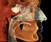
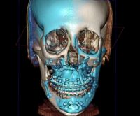
- Using a single scan to capture a 3D image of teeth, roots, jaw problems and sinuses without distortion of the picture.
- Allowing dental professionals to see exact tooth position and relationship of anatomy
- Providing a lower dose detailed scan while decreasing the radiation risk
- Providing adaptable settings to allow professionals to select customized fields of view for targeted areas of interest
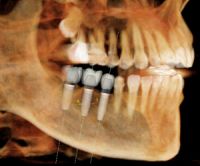
- Allowing dental experts surgical predictability with treatment planning in 3D with restorative outcomes. Ther allows the doctor to map out the entire treatment course from implant placement to final restoration
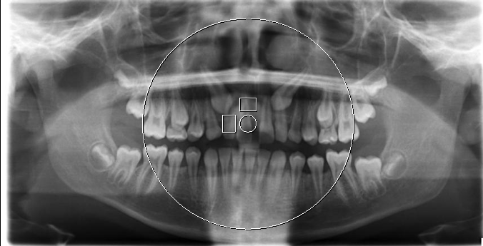
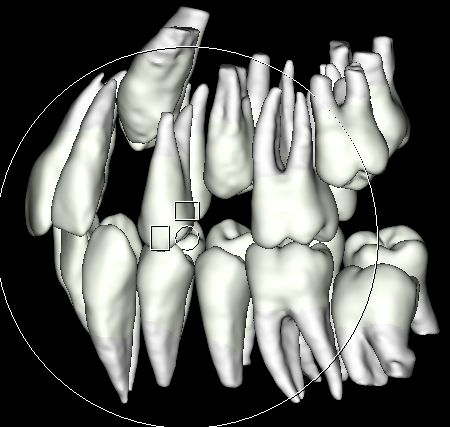
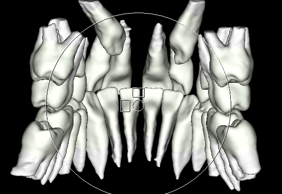
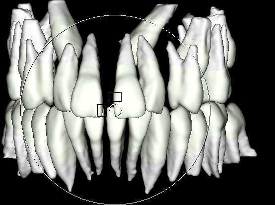
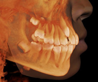
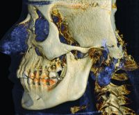
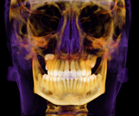
Why we use 3D imaging?
Case example of bilaterally impacted canines (i-teeth). Notice in the 2D xray it is impossible to see where in the jaw they are positioned, if they are savable, and the best route to bring them in. The same patient with 3D imaging reconstructed by Dr. Spector revealed the precise position of the impacted teeth within bone. Benefits: safer, less trauma, higher success
Dental professionals interested in referring patients for the i-CAT scan, click the refer button below