Advanced 3D Imaging Streamlines Treatment For Patients
ROUND LAKE, Ill.- Remarkable expertise combined with advanced technologies leads to outstanding orthodontic care. That’s exactly what patients experience at Stosich Consulting. Dr. Michael Stosich incorporates 3D image-guided orthodontics into the diagnostic process.
A thorough understanding of each patient’s orthodontic situation and a proper diagnosis is key to achieving optimal results. Our patients benefit from the 3D pictures provided by advanced imaging technology. Rather than producing a stagnant 2D image which fails to communicate precise tooth placement and depth, we can gain a stunningly clear 3D view of teeth and the jaw, and even the entire skull, when necessary.
“The insight 3D imaging provides assists me in accurately diagnosing problems. This information is critical in determining the best method of treatment and creating an appropriate treatment plan for patients,” says Dr. Stosich, an award-winning orthodontist in the Round Lake area.
When performing 3D image-guided orthodontic diagnostics, we digitize our patients dental molds and scan the area of interest. The result is a detailed scan of the entire mouth that is captured and viewable on a computer screen. This way, patients can get an in-depth view of their oral anatomy and a better understanding of their dental and orthodontic health.
Advantages of 3D imaging include:
- Dr. Stosich can view exact tooth placement in relationship with each patient’s unique oral anatomy.
- One image can capture teeth, roots, jaw bones and sinuses in their entirety with amazing clarity.
- Adjustable settings allow Dr. Stosich to customize images according to targeted areas of interest.
- Scans with low radiation dosages keeps patients safe.
- Detailed 3D images give Dr. Stosich enhanced surgical predictability in planning care.
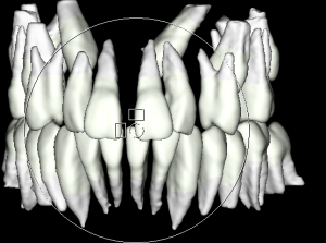
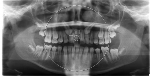
Check out the below photos that feature bilaterally impacted canines on the same patient. On the left is a 2D image, which doesn’t offer a clear indication of the condition of teeth, jaw placement or a good route to bring the teeth in. The right picture, however, is a 3D image of the patient. Here, we can see the exact position of the impacted teeth.
3D technology is accessible to a handful of orthodontics practices, but not all are equipped with the skill set to translate these technologies into improved patient comfort and optimal results. Dr. Stosich, who attended top universities and has received twice the necessary training to practice orthodontics, has years of experience harnessing the best industry technologies to streamline the treatment process and achieve the best possible outcome for his patients. The proof is in his credentials- Dr. Stosich also serves as the attending orthodontist for the University of Chicago Medical Center’s craniofacial and cleft palate team.
To learn more about Dr. Stosich, 3D imaging or the safety precautions we practice when taking X-ray images, contact our office. Stosich Consulting is a trusted source of dentofacial orthopedics, Invisalign and braces in the Round Lake, Grayslake, Kenilworth, Skokie and Wilmette areas.